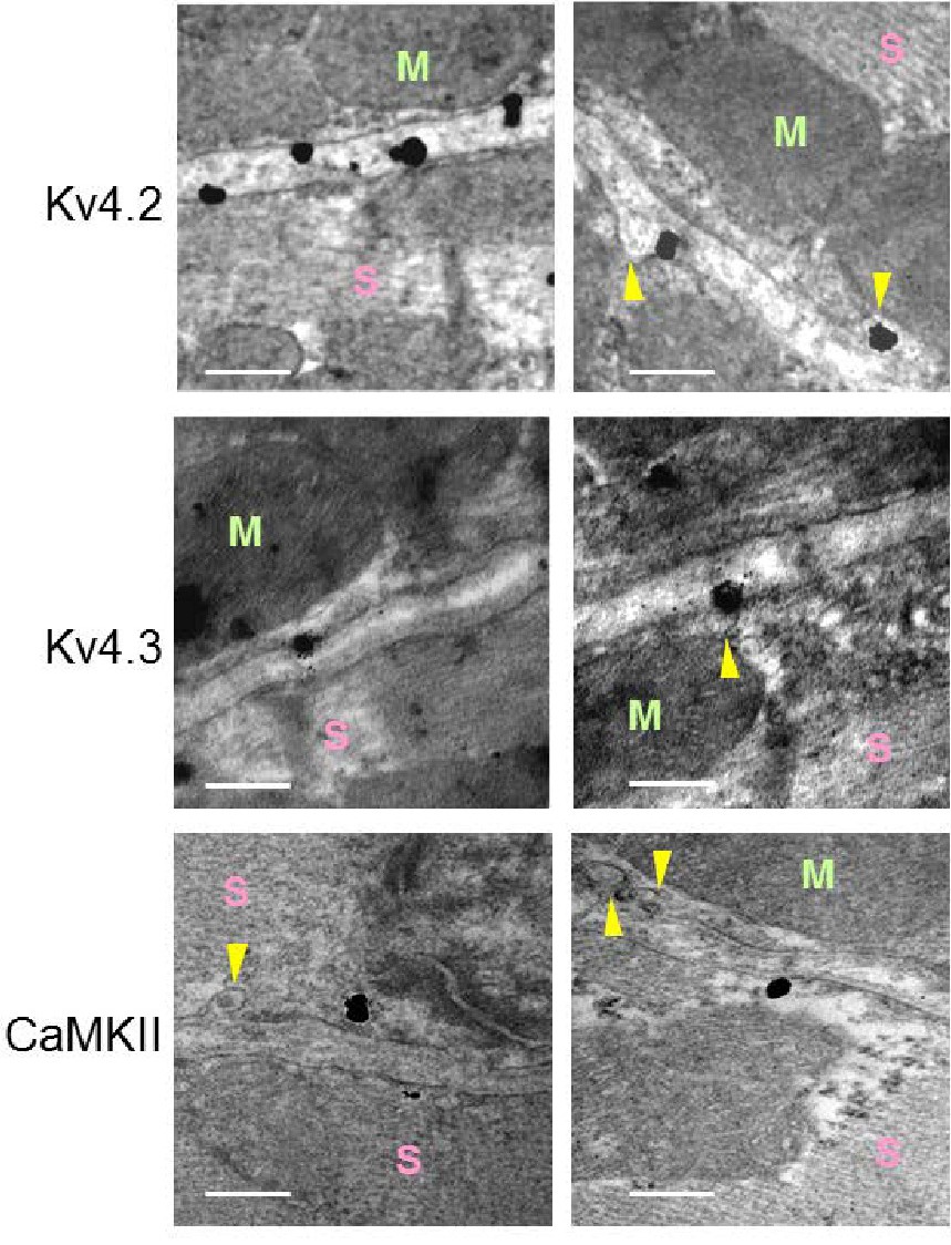
Fig. 4. Kv4.2 and Kv4.3 locate in caveolae and in planar membrane, whereas CaMKII only in non-caveolar regions. Caveolae (yellow arrowheads) were identified because of its flask shaped structure. Silver-enhanced immunogold staining showing Kv4.2 and Kv4.3 proteins in caveolae and in planar regions of the membrane, and CaMKII only in non-caveolar regions. Representative electron microscope images of 4 rat ventricles (Calibration Bar = 200 nm; M = Mitochondria; S = Sarcomere).